diagnostic testing

X-Ray images are a quick, cost effective, and excellent tool for screening for fractures and dislocations. They are widely available in orthopaedic offices. They are also very efficient as a follow-up tool to ensure proper healing.
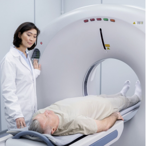
Computerized Tomography (CT) scans are available in the hospital and provide a quick imaging tool which provides excellent 3D bone imaging. They can detect subtle fractures in the bone and are helpful to a surgeon in pre-operative planning in complex cases.
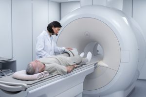
Magnetic Resonance Imaging (MRI) Scans provide exceptional images and are the best choice for viewing soft tissues. However, the scan is lengthy and more expensive than other imaging techniques.
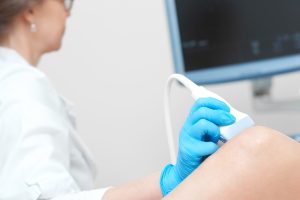
Ultrasound uses sound waves to create an image. This type of imaging is fast, inexpensive, and uses no radiation. The operator can take 2D pictures with it but real time imaging is the most helpful. It can be used to image fractures, check bone healing, and see tendon/ligament injuries.
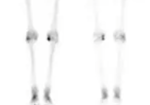
A Bone Scan is a type of nuclear imaging where a small amount of radioactive tracers are introduced into the body and photographed with a special camera over the course of several hours. The tracers collect in areas of the bone that have a different metabolism due to injury, infection, or disease. This test is helpful when there are unexplained reasons for bone pain.
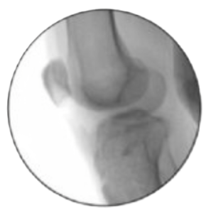
Fluoroscopy imaging provides a real time, low dose x-ray image in 2D or 3D. The images are not as crisp as traditional x-ray, but it is portable and allows an orthopaedic surgeon to do quick progress checks on implants during surgery.
